


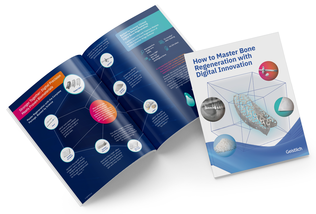


Why Clinicians Choose Palate Free Treatments

Non-Inferior to Gold Standard
Geistlich Fibro-Gide® offers a reliable, non-inferior alternative to CTG in augmenting soft tissue around implants1

Time
Saves up to 7 minutes of chair time on average1

Fewer Surgical Procedures
Eliminates the need to harvest an autologous graft
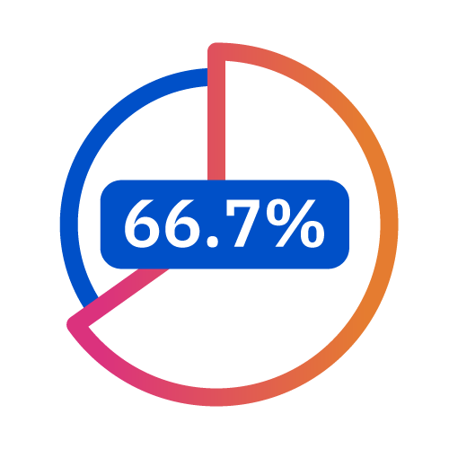
Patient Satisfaction
66.7% of patients prefer Geistlich Fibro-Gide® over CTG2

Begin with a choice
What is the right choice for you?
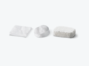
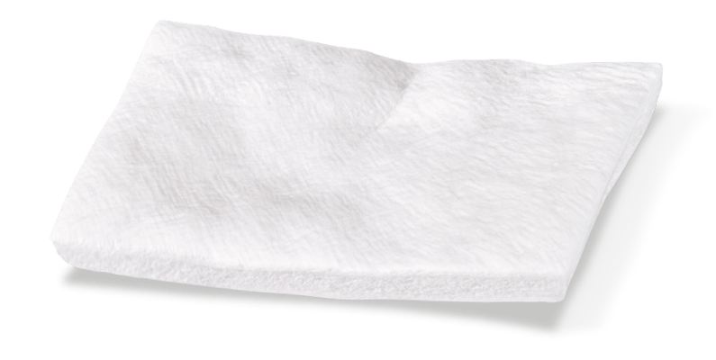
Geistlich Mucograft®
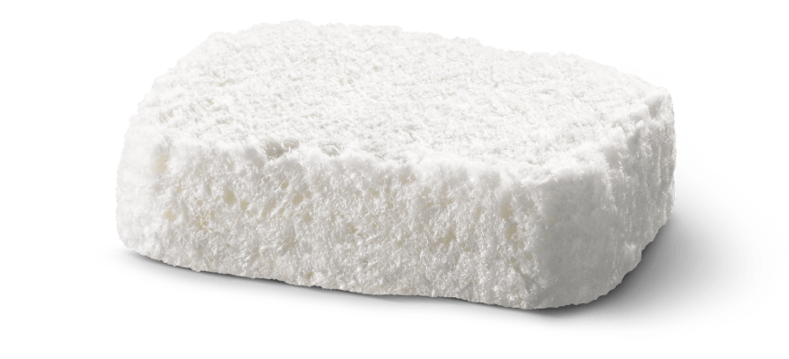
Geistlich Fibro-Gide®
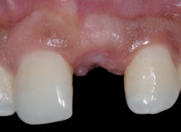
Socket Seal
Vestibuloplasty
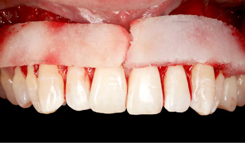
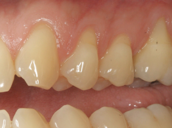
Recession Coverage
Outcomes
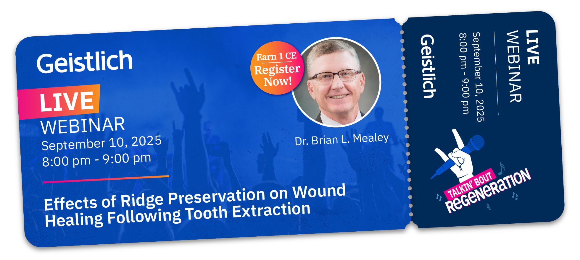
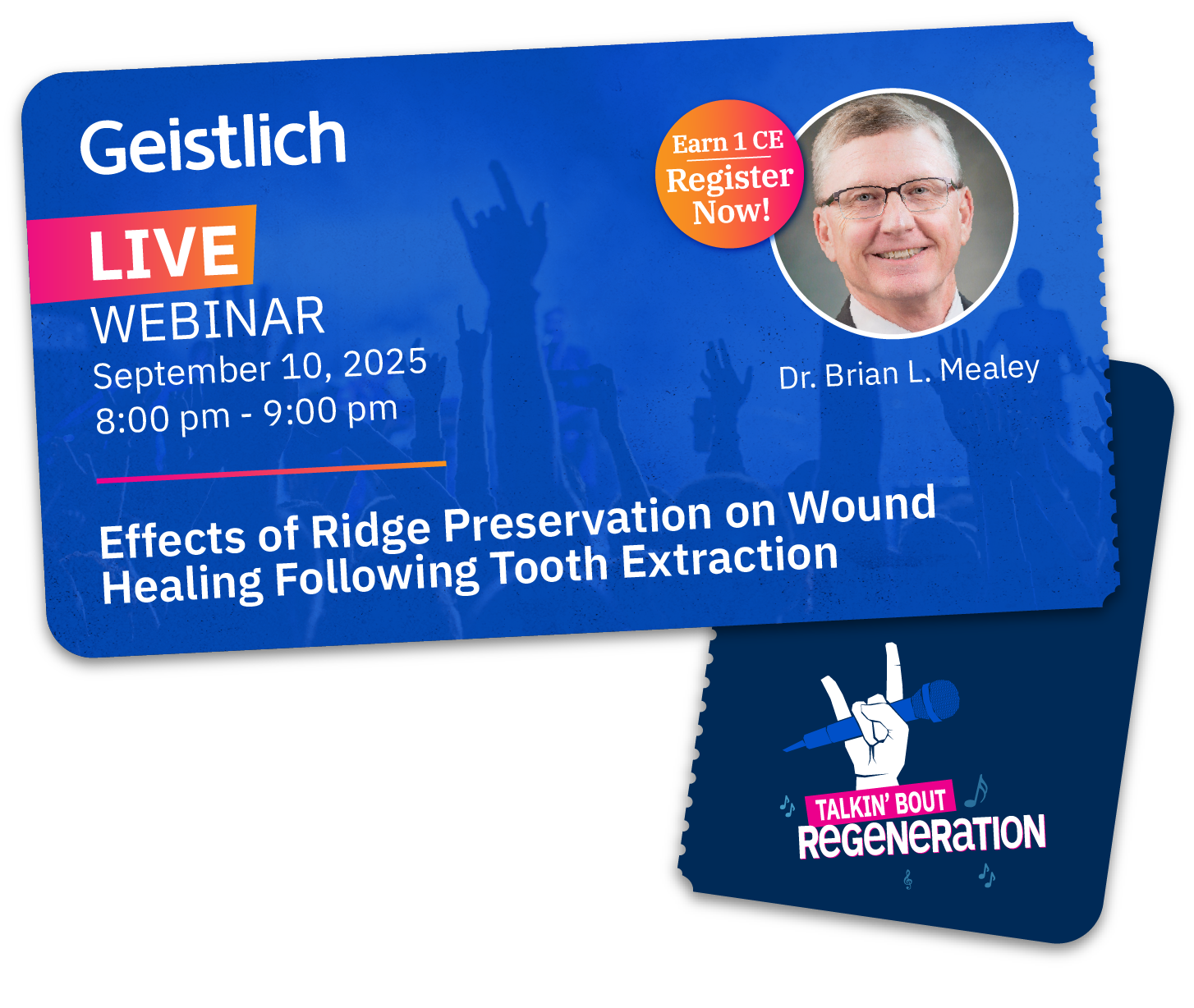
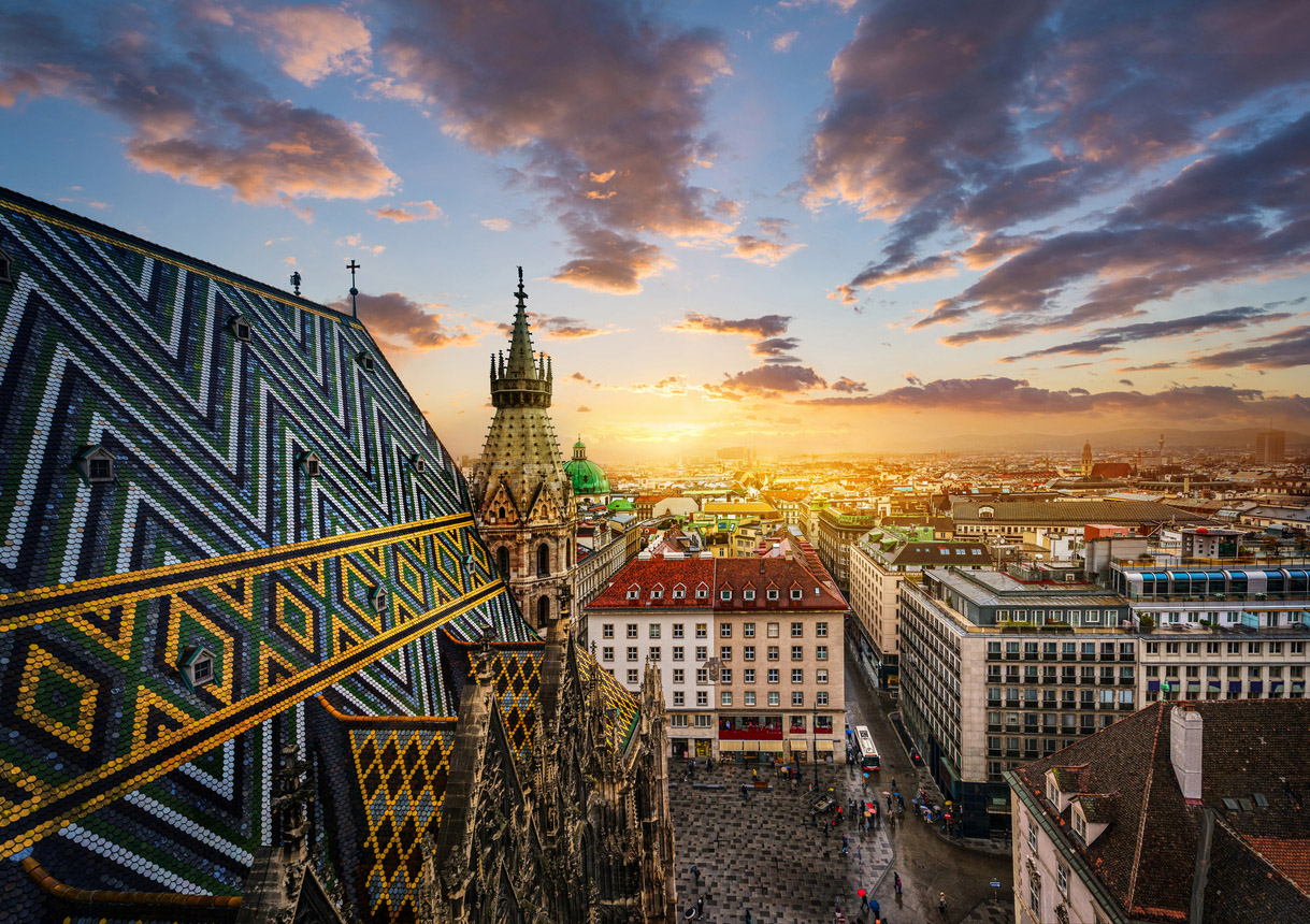
![]() Geistlich Education Sweepstakes:
Geistlich Education Sweepstakes:
Attend & Win a Trip to the International Osteology Symposium – Vienna 2026!
Explore all webinars and a chance to win a trip to Osteology 2026 in Vienna
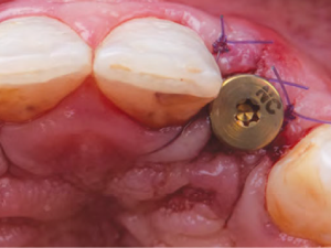
 CASE STUDIES
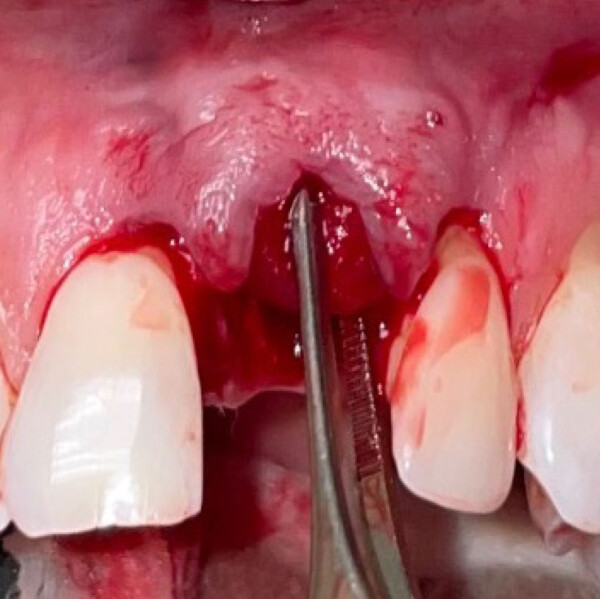
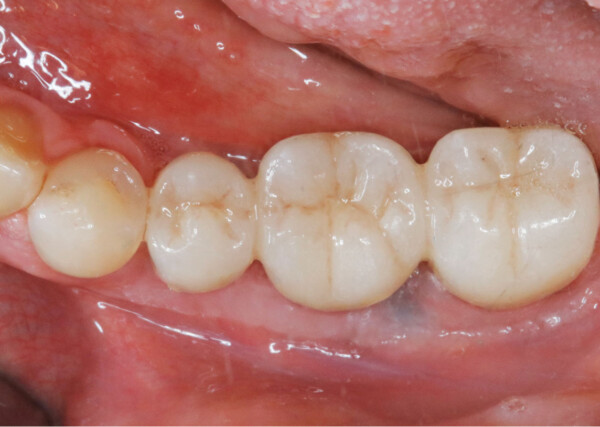
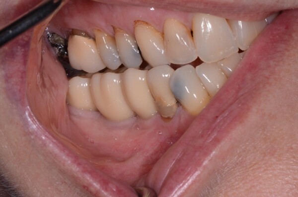
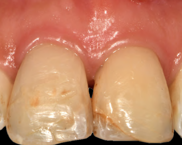
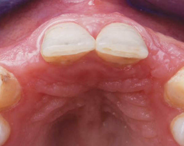
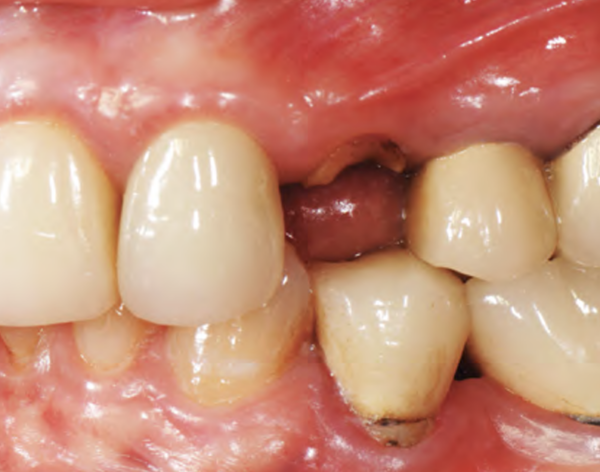
Our Palate Free Approaches
to Soft Tissue Treatment
 Clinical Videos
Geistlich Fibro-Gide® vs Geistlich Mucograft®
Soft Tissue Thickening with Geistlich Fibro-Gide®
Recession Coverage with Geistlich Fibro-Gide®
Surgical Videos
Root Coverage of a Single Recession Defect
Soft-Tissue Augmentation around Dental Implants – Case 1
Soft-Tissue Augmentation around Dental Implants – Case 2
Handling Videos
Geistlich Fibro-Gide® Introductory Training
Handling Geistlich Mucograft®
Handling Geistlich Mucograft® Seal
- Clem et al., J Periodontol. 2023;1–10.
- McGuire, M. et al. (2022). JPeriodontol. 93(3): 333-342.
- McGuire, M.K. et al. (2014). J Periodontol. 85(10):1333-41
- Thoma DS. et al. J Clin Periodontol. 2020 May;47(5):630-39
